
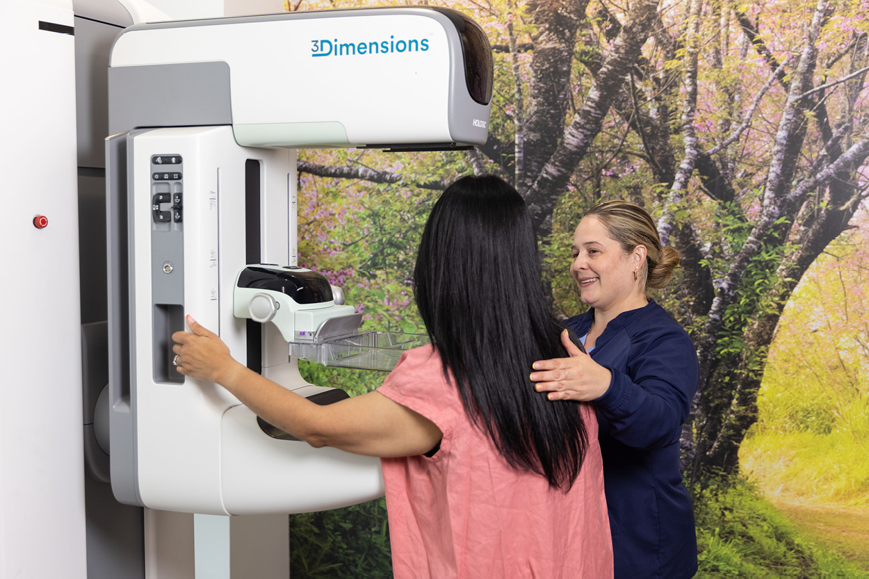
When Mammograms Need a Closer Look
Most women who have a mammogram are told that everything looks normal. A smaller number get a call from our team because our radiologist noticed something that needs another look. Maybe it’s a small cluster of calcium deposits, a mass with an uneven edge, or an area of tissue that appears denser than before. Getting that call can be unsettling, but follow-ups like this are common.
At NY Imaging Specialists, we offer screening mammograms before any symptoms appear so we can catch changes in breast tissue early, while they're still easiest to address. Most guidelines suggest starting yearly mammograms at age 40, but your doctor might recommend a different schedule based on your family history or other risk factors. Each year, our radiologist compares your new images against your prior ones, looking for anything that's shifted since your last scan. A single image only shows what's there now. Comparing it to last year's is what lets us catch a change before it becomes something more significant.
Screening Versus Diagnostic
Your first mammogram is a screening exam, done on schedule and without symptoms, to check your breast health before anything feels wrong. If our radiologist notices something, any follow-up imaging is called “diagnostic”. That single shift from screening to diagnostic changes the exam's purpose entirely. A screening mammogram looks broadly for anything unfamiliar. A diagnostic exam is narrower and more deliberate, built around one specific area and one specific question, using more detailed views to get an answer a screening exam wasn't designed to give. That difference in purpose, not the gravity of what was found, is why a callback happens. It does not mean something is wrong. It means one area needs a more focused look than a routine scan can provide.
When our radiologist asks for a follow-up, they describe what they see, rather than forming a diagnosis. That description needs more information before our team can interpret it, and diagnostic imaging provides it. What happens next depends on what the original mammogram showed. A diagnostic mammogram takes additional, more detailed images of the specific area in question. Our radiologist may also order an ultrasound to determine whether an area is solid or fluid-filled, since a cyst and a solid mass can look similar on a mammogram but appear very different on an ultrasound. An MRI is used less often, typically when someone has dense breast tissue that makes mammograms harder to read, or when a more detailed, three-dimensional view is needed. On the rare occasion that diagnostic imaging still leaves a question unanswered, a biopsy may be the next step to get a definitive answer, though that's a separate conversation your doctor will walk you through if needed.
The Anxiety is Real
Getting called back can bring on a wave of worry, and there's nothing unreasonable about feeling that way. Most callbacks end with a diagnostic mammogram or ultrasound that confirms the area is benign, a cyst, dense but normal tissue, or simply a finding that needed a clearer picture to explain. If you have questions before your appointment, our team is here to walk you through what to expect so you're not sitting with uncertainty longer than you need to.
At NY Imaging Specialists, your diagnostic follow-up happens in the same comfortable, private setting as your original screening, with a radiologist who already knows exactly what to look for. The sooner that appointment happens, the sooner you trade the confusion for an answer.